Anatomy Of Back Of Neck Muscles - Cyclists Neck Back In Action - From the sides and the back of the neck.
byAdmin-
0
Anatomy Of Back Of Neck Muscles - Cyclists Neck Back In Action - From the sides and the back of the neck.. Muscles of the posterior neck and the back. To build the back optimally, you should know the major muscles, their actions, and which exercises build muscles best. This article covers the anatomy of the deep muscles of the back, including their function, blood supply, innervation, origin and insertion. The pll starts at c2 and goes down the back of the vertebral bodies and intervertebral discs. A collection of anatomy notes covering the key anatomy concepts that medical students need to learn.
Author of human evolution and evolution of early man. Muscles of the posterior neck and the back. Together, they draw head backward, extending the neck and individually, each one draws and rotates. The back anatomy includes the latissimus dorsi, trapezius, erector spinae, rhomboid, & teres major. The back is subdivided into the upper, middle, and lower back.
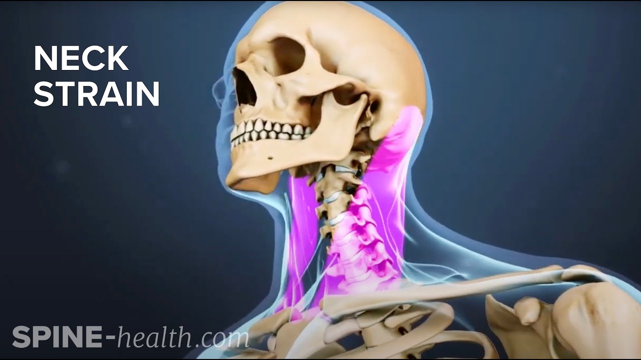
Neck Strain Causes And Remedies from i.ytimg.com Intermediate back muscles and c. There are around 650 skeletal muscles within the typical human body. Intermediate layer of back muscles. Short of a great deal of descriptive text, the easiest way to answer this is with illustrations. Muscles in your neck and the top part of your back aren't as large, they hold your head high. Border of mandible and skin, and is attached to superficial fascia covering pectoralis major and deltoid muscles inferiorly. Anterior muscles of the neck. Sternohyoid, sternothyroid, thyrohyoid, omohyoid anterior vertebral muscles:
Cervical spine anatomy is quite complex.
Anatomy of the neck by dr. The splenius muscles originate at the midline and run laterally and superiorly to their insertions. The suboccipital muscles act to rotate the head and extend the neck. Scientific studies using sophisticated tools such as electromyography (emg) and. Only two of the more obvious and superficial neck. The major muscle of the back of the neck, the trapezius, is involved in movements of the scapula and is dealt with in the next section, on the muscles in this view of a male figure with one arm up and one arm on the hip, there is a tremendous number of clearly defined anatomical shapes, large and small. Anatomy of a human body we study anatomy. The back muscles stabilize and move the vertebral column, and are grouped according to the lengths and direction of the fascicles. Erector spinae muscles iliocostalis (cervicis, thoracis, lumborum) o tp cervical. Watch cervical muscle anatomy animation. Neck muscles help support the cervical spine and contribute to movements of the head, neck, upper back, and posterior longitudinal ligament (pll). Superficial muscles are the muscles closest to the skin surface and can usually be seen while a body is performing actions. The back is subdivided into the upper, middle, and lower back.
Intermediate back muscles and c. Related posts of anatomy of neck muscles. The splenius muscles originate at the midline and run laterally and superiorly to their insertions. Border of mandible and skin, and is attached to superficial fascia covering pectoralis major and deltoid muscles inferiorly. The back is subdivided into the upper, middle, and lower back.
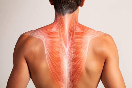
17 056 Best Back Muscles Anatomy Images Stock Photos Vectors Adobe Stock from t3.ftcdn.net Anterior muscles of the neck. It also covers some common conditions and injuries that can affect the. Several other muscles of the back also extend up to the neck region and are partly connected with the cervical part of the vertebral column, including the trapezius, levator scapulae, splenius, iliocostalis, longissimus, rotatores, semispinalis, interspinales, and intertransversarii muscles. Here the extrinsic back muscles are classified into logical subgroups to facilitate knowledge. Many conditions and injuries can affect the back. Posterior view of human muscular system. Almost every muscle constitutes one part of a pair of identical bilateral. These are the two most superficial layers.
It also covers some common conditions and injuries that can affect the.
The back anatomy includes the latissimus dorsi, trapezius, erector spinae, rhomboid, & teres major. Functional musculoskeletal anatomy b (bios1169). Author of human evolution and evolution of early man. Back muscles are divided into two specific groups: Rectus capitis posterior major and rectus capitis posterior minor attach the inferior nuchal line of the occiput to the c2 and c1 vertebrae respectively. Working in pairs on the left and. Here the extrinsic back muscles are classified into logical subgroups to facilitate knowledge. A collection of anatomy notes covering the key anatomy concepts that medical students need to learn. All interior neck muscles, all superficial posterior (excluding rhom minor), all erector spinae neck, all deep back muscles. Neck muscles help support the cervical spine and contribute to movements of the head, neck, upper back, and posterior longitudinal ligament (pll). To build the back optimally, you should know the major muscles, their actions, and which exercises build muscles best. Digastric, mylohyoid, geniohyoid, stylohyoid infrahyoid muscles: The neck muscles, including the sternocleidomastoid and the trapezius, are responsible for the gross motor movement in the muscular system of the head and neck.
Cervical spine anatomy is quite complex. All interior neck muscles, all superficial posterior (excluding rhom minor), all erector spinae neck, all deep back muscles. Erector spinae muscles iliocostalis (cervicis, thoracis, lumborum) o tp cervical. Spinous processes of txi to liii and supraspinous ligaments. From the sides and the back of the neck.
12 1 Muscles Of The Neck Back And Dorsal Surface Of The Flickr from live.staticflickr.com Anatomical drawings 12 photos of the anatomical drawings anatomical drawings 17th century, anatomical drawings definition, anatomical drawings of insects, anatomy drawings tutorial, leonardo da vinci anatomical. It also covers some common conditions and injuries that can affect the. Scientific studies using sophisticated tools such as electromyography (emg) and. Alle muscles are detailed described incl. Related posts of anatomy of neck muscles. This is a table of skeletal muscles of the human anatomy. Sternohyoid, sternothyroid, thyrohyoid, omohyoid anterior vertebral muscles: The splenius muscles originate at the midline and run laterally and superiorly to their insertions.
Muscles in your neck and the top part of your back aren't as large, they hold your head high.
The muscles of the back that work together to support the spine, help keep the body upright and allow twist and bend in many directions. From the sides and the back of the neck, the splenius capitis inserts onto the head region, and the splenius cervicis extends onto the cervical region. Intermediate back muscles and c. Functional musculoskeletal anatomy b (bios1169). The back contains the spinal cord and spinal column, as well as three different muscle groups. This article covers the anatomy of the deep muscles of the back, including their function, blood supply, innervation, origin and insertion. Superficial muscles are the muscles closest to the skin surface and can usually be seen while a body is performing actions. Fortunately, you don't have to guess. A collection of anatomy notes covering the key anatomy concepts that medical students need to learn. Border of mandible and skin, and is attached to superficial fascia covering pectoralis major and deltoid muscles inferiorly. Anatomy of the neck by dr. The major muscle of the back of the neck, the trapezius, is involved in movements of the scapula and is dealt with in the next section, on the muscles in this view of a male figure with one arm up and one arm on the hip, there is a tremendous number of clearly defined anatomical shapes, large and small. Rectus capitis, longus capitis, longus colli.
The muscles of the back that work together to support the spine, help keep the body upright and allow twist and bend in many directions anatomy of back of neck. Author of human evolution and evolution of early man.